 |
| Work at Sea...Techniques |
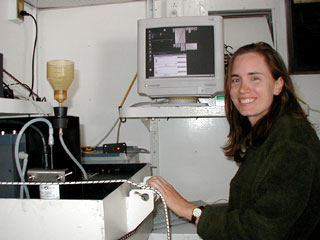
| Flow cytometry is used to measure the optical properties of cells in a flow stream. Since plankton are naturally suspended in sea water, flow cytometry is a good way to study them. The FlowCAM is an instrument that uses flow cytometry to count and image cells with photosynthetic pigments. It is able to do this by detecting fluorescence. At each station, we help Nicole use FlowCAM to study plankton populations between 5 and 200 micrometers in size. We look at water samples collected from various depths. We are able to study phytoplankton and even some zooplankton. |
 |
|
|
|
|
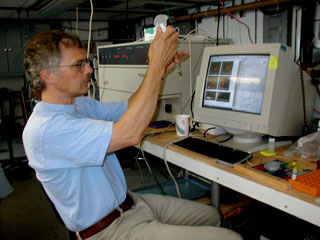
Another important on-board activity is quantifying the amount of chlorophyll in the water column. Water samples are filtered to concentrate cells into two size classes – less than 20 micrometers and less than 3 micrometers. Then we help Renee soak the filters in acetone to extract the chlorophyll from the cells. We expose the acetone solution to a beam of blue light that excites the chlorophyll within the solution and causes it to emit red light. We use the red light emission to quantify the amount of chlorophyll in each sample. |
|
|
|
|
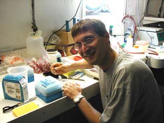
Staining water samples and making slides allows us to actually see (with the aid of a microscope) the very tiny cells we have been studying. We help Polly add stains to filtered water samples (the samples were filtered to remove cells greater than 20 micrometers). We add different stains that bind to specific parts of the cells; one stains DNA and another stains cytoplasm. We then filter the stained samples through a black membrane filter that allows the water to pass through but catches the cells. The membrane filter is placed on a microscope slide and topped with a drop of oil and a cover slip. Since these slides are not
transparent, the microscope illuminates them from above rather than
below. Different types of light are used to excite the differentially
stained materials. |
| Another technique that uses flow cytometry is the FACScan. The FACScan is designed to count (but not image) smaller plankton cells. We help Ed use the FACScan to count bacteria and help determine what percentage of the bacteria is active. To do this, we have to stain the samples before running them on the FACScan. We stain them with a chemical that indicates which bacteria are actively respiring. In addition to counting bacteria, we also help Ed use the FACScan to count larger cells (up to 15 micrometers). We can use this information to help characterize the biological cycles where bacteria thrive. |
 |
| In order to directly measure bacterial growth rates, we help the scientists use radioactive tracers. We introduce a radioactive substance into filtered water samples (the samples were filtered to remove cells larger than bacteria) and then let the samples incubate. During the incubation period, active cells incorporate the tracer into their proteins. We are then able to measure the amount of radioactivity in the cells and from that calculate the percentage of active cells. |
|
|
| One way to study the diversity of marine bacteria in the water column is to look at genetic similarities and differences between types of cells. We help Andrew collect DNA from the bacteria in the water samples. After we collect the DNA, we will use the information to study the marine bacterial community. |
 |
|
|
To look at the viruses that are present in the water column, we help David stain the water samples with a chemical that binds to nucleic acids (DNA or RNA). We then filter the water to only allow cells smaller than 0.2 micrometers through. We then analyze the stained viruses under fluorescent light with a high-powered microscope.
Dissolved organic carbon (DOC) plays an important role in the marine carbon cycle (link to nutrient cycling). We help the scientists measure the amount of DOC in the water samples by heating the samples to 680°C (1256°F) and measuring the amount of carbon dioxide gas emitted. |
|
|
| <<<Look Back |